Research Infrastructure

Methods

Nuclear Magnetic Resonance

NMR Spectroscopy

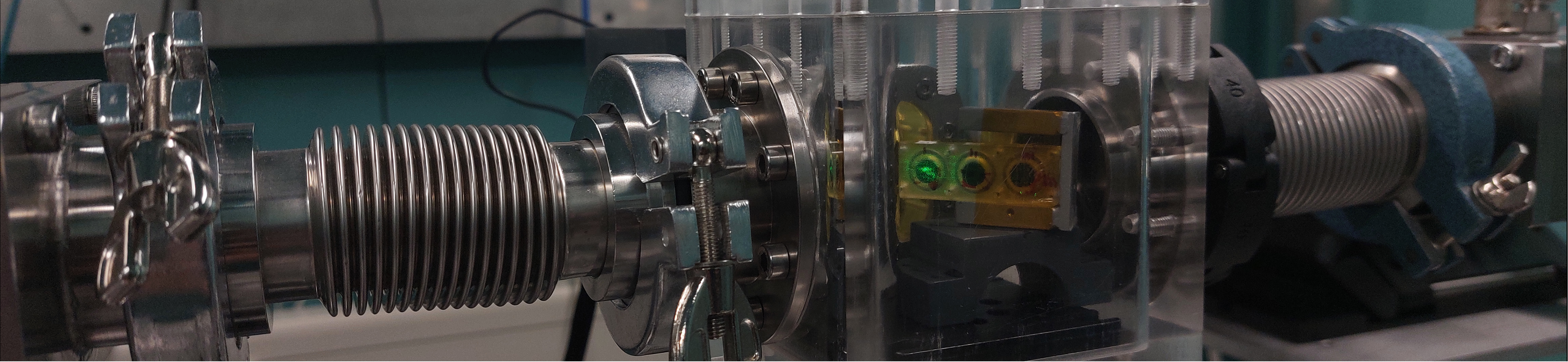
NMR spectroscopy is one of the most powerful analytical techniques available to the preparative chemist. Many nuclei are NMR-active and exhibit chemical shifts that characteristically reflect their molecular environment. With the availability of a 400 MHz NMR spectrometer directly in the radionuclide-approved laboratories at KIT-INE, multi-nuclear NMR spectroscopy is routinely used to study reactions and reaction products of technetium. Methods include NMR directly at the Tc-99 nucleus, as well as 2D experiments and isotopic labelling strategies for the observation of technetium-bound nuclei with low abundance. In combination with other analytical techniques, NMR spectroscopy often allows an unequivocal structural assignment in solution even when no single-crystal diffraction data are available.

Single-Crystal X-ray Diffraction

Crystallography

Single crystals consist of an asymmetric unit organized in an often periodic crystal lattice. The asymmetric units exhibit long-range ordering consistent with the symmetry requirements of a specific space group. Due to the periodic repeating arrangement in most single crystals, they interact with X-ray photons of a wavelength similar to the interatomic distances (about 1-2 Å), resulting in constructive or destructive interference causing a diffraction pattern. From this pattern, the molecular arrangement of the compound forming the crystalline material can be determined as a definite, unambiguous proof of its structure in the solid state. Crystallographic results are evaluated in conjunction with solution-state methods such as multinuclear NMR spectroscopy or ATR-FT-IR spectroscopy.


Advanced X-ray absorption spectroscopy

EXAFS & XANES

Upon interaction with high-energy photons in the correct energy range, the inner-shell electrons in close proximity to the atomic nuclei can be ejected, resulting in ionization and the formation of an inner-shell electron hole. The absorption of the photons but also the emission of X-ray fluorescence as a consequence of the relaxation of a valence electron down to the generated core hole are element specific — their energy corresponds to the energy difference between the electron shells. The probing of these interactions of X-ray photons with matter is X-ray absorption spectroscopy (XAS). However, the measured signals have a fine structure that is revealed when brilliant light sources (e.g., synchrotron radiation) are combined with highly sensitive detectors. The fine structure reveals neighboring atoms in the near-field of the targeted nucleus. Such features can be analyzed by evaluating the near-edge region — X-ray absorption near edge spectroscopy (XANES) — or in the resonant region beyond the absorption edge — extended X-ray absorption fine structure (EXAFS) analysis. Advanced X-ray absorption spectroscopy thus complements diffraction-based methods that require long distance ordering. Normally, measurements are performed at the K-edge of technetium because the required 21 keV photons are readily available. KIT-INE offers a unique opportunity for measurements at the relatively low energy L3-edge of technetium (ca. 2.7 keV). Due to the proximity of the L3 shell to the valence electrons, the spectrum is much more influenced by the chemical environment of technetium compared to the K-edge.

Attenuated total reflectance (ATR)

Infrared Spectroscopy

Chemical bond vibrations are induced upon the interaction of molecules with photons from the low-energy regime of the electromagnetic spectrum. Characteristic molecular vibrations can be diagnostic for specific functional groups. Vibrations follow selection rules derived from symmetry considerations in line with the point group of the molecule. Generally, vibrations that significantly change the dipole moment of a molecule are well-suited for analysis by infrared (IR) spectroscopy. For example, nitrosyl and carbonyl ligands show a prominent valence vibrational band around 1800 cm-1 and 2000 cm-1, respectively. The original sweeping of the IR range in such spectrometers was achieved by a continuous variation of the energy of the incident light, e.g., through modulation and selection by a grating monochromator. However, modern spectrometers use interferometers (e.g., Michelson interferometer) and a reference laser. After the Fourier transformation (FT), the signals are transferred into the desired wavenumber space. The main benefits of FT-IR spectroscopy are an unmatched acquisition speed and dynamic resolution. The standard sampling mode is the transmission of an IR beam through the sample. However, attenuated total reflectance (ATR) is a modern variant that allows the sampling of extremely small sample volumes by measuring the reflectance change at the surface of an ATR crystal (e.g., diamond, Si, Ge). The group operates several FT-IR spectrometers with different ATR units inside the radioactive laboratories at KIT-INE.